[en] The SARS-CoV-2 is a 30kb-long single-stranded positive sense RNA virus. It evolves due to host antiviral defenses, mainly driven by APOBEC deaminase activity. APOBEC3 enzymes play a crucial role in restricting viruses by introducing mutations into their genome. These mutations occur in specific contexts, specifically when cytidine is preceded by thymine or uracil, known as the 5’TC or 5’UC motif. The selective pressure exerted by APOBEC3 enzymes results in a depletion of these motifs in exposed viruses, a phenomenon termed the "APOBEC3 footprint."
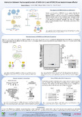
Viruses have evolved various mechanisms to antagonise the APOBEC3 response or even to exploit it to promote genetic diversification of the viral genome. Antagonism relies on the use of viral proteins that recruit APOBEC proteins and target them for degradation or delocalisation away from the viral replication centre. We therefore proposed to generate an interactome map of SARS-CoV-2 proteins with different APOBEC proteins using high-throughput GPCA assays. We identified an interaction between APOBEC3G (A3G) and APOBEC3H (A3H) with Nucleocapsid (N).
To better decipher this interaction, we tested which domain of each protein are involved. We performed a GPCA assay and found that the N CTD domain (aa 255-365) and the A3G CD1 domain (aa 1-191) are involved in the interaction between these proteins. Both are RNA binding domains, so RNA may be involved in this interaction. To test this hypothesis, we performed a Co-IP in HEK-293T cells in presence or absence of RNase.
We finally wanted to confirm the physiological interaction between A3G and N during SARS-CoV-2 infection. To this end, we performed a Co-IP in HBEC-3KT cells stably expressing ACE2 and TMPRSS2 infected with the SARS-CoV-2 Wuhan strain. Initial results indicated an interaction between A3G and N, prompting ongoing investigations into the impact of this interaction on the potential antiviral activity of A3G.