[en] Impact of neuroinflammation developed during experimental autoimmune encephalomyelitis on hippocampal synaptic plasticity and cognition
Recent evidence clearly showed that the immune system plays an important role on many neuronal functions like cognition and synaptic plasticity. A complex crosstalk takes place between neurons, astrocytes, microglia and infiltrating immune cells to promote the remodeling of synaptic circuits. Glial cells are now considered as the third element of synapses contributing to neurotransmission and neural plasticity. However, this system can be rapidly disrupted during inflammatory conditions leading to cognitive impairments. These deficits are very common in neuroinflammatory disorders but the mechanisms involved are still poorly understood. Immune responses are very large and complex, being either neuroprotective or detrimental, and are dependent on the context, the duration of the inflammatory process and the type of activated inflammatory cells.
This project aims to study the effects of neuroinflammation on neuronal network activity and synaptic plasticity in mouse hippocampus and to highlight the molecular and cellular inflammatory actors related to cognitive disorders. We use EAE (experimental autoimmune encephalomyelitis), a model of MS, as a model of CNS chronic inflammatory disease that we induce by a specific autoimmune reaction leading to demyelination and motor disorders. EAE develops with a relapsing-remitting course allowing us to analyse the different inflammatory steps and their impact on cognition.
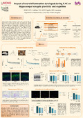
Hippocampal synaptic plasticity was analyzed during the course of EAE by ex vivo electrophysiological recordings (LTP) made on acute hippocampal slices from EAE mice. LTP measurements showed that the level of potentiation is higher at the peak of EAE but progressively decreases during the remission phase to reach a level significatively lower than the control one suggesting a time dependent impairment of hippocampal plastic potential during the EAE remission stage. Spatial learning of remitting mice was evaluated in vivo by the contextual fear conditionning and revealed a cognitive impairment during the remission stage.
Although myelin is the main target of the immune reaction during EAE, no modification of MBP expression was found by western-blotting and IHC in mouse hippocampus at any stage of EAE. Despite the lack of demyelination, our immunostainings and ELISA experiments revealed a higher glial activation and a production of inflammatory factors like IL1beta in the hippocampus of EAE mice. The number of both astrocytes and microglial cells follows the disease progression as it enhances at the peak of the disease and then decreases during the remission stage.
So, our results demonstrate that immune responses and neuroinflammation developed during EAE can also affect cognitive structures like hippocampus and can lead to cognitive impairments. As no demyelination occurs, activated microglia and astrocytes could be linked to modifications of hippocampal synaptic plasticity during EAE and could therefore be important actors implicated in cognitive disorders related to neuroinflammation.